Heads or tails? Scientists identify gene that regulates polarity in regenerating flatworms

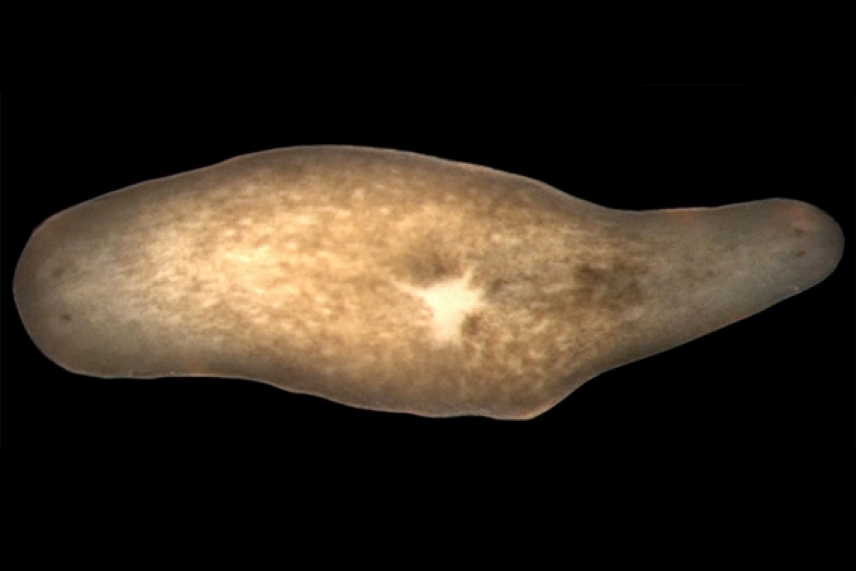
Inhibition of the gene beta-catenin causes worms to regenerate a head instead of a tail (right head).
Image: Christian Petersen
CAMBRIDGE, Mass. - When cut, a planarian flatworm can use a population of stem cells called neoblasts to regenerate new heads, new tails or even entire new organisms from a tiny fragment of its body. Mechanisms have been sought to explain this process of regeneration polarity for over 100 years, but until now, little was known about how planaria can regenerate heads and tails at their proper sites.
Scientists in the lab of Whitehead Member Peter Reddien have discovered that the gene Smed-beta-catenin-1 is required for proper decisions about head-versus-tail polarity in regenerating flatworms. Their results were published in the December 6 issue of Science Express online.
Reddien’s lab studies regeneration in the planarian Schmidtea mediterranea. “Evolution has selected for mechanisms that allow organisms to accomplish incredible feats of regeneration,” and planaria offer a dramatic example, notes Reddien, who is also an assistant professor of biology at Massachusetts Institute of Technology. “By developing this model system to explore the molecular underpinnings of regeneration, we now have a better understanding of the process.”
The researchers used a technique called RNA interference (RNAi) to screen a group of genes known to be involved in animal development, in order to study the signaling mechanisms that regulate whether the animal would produce a head or tail during regeneration.
“We discovered that inhibiting the gene Smed-beta-catenin-1 caused animals to regenerate a head instead of a tail at the site of the wound,” says Christian Petersen, Whitehead postdoctoral fellow and lead author on the paper. “This resulted in a worm that possessed two oppositely facing heads. Smed-beta-catenin-1 is the first gene found to be required for this regeneration polarity.”
Genes very similar to Smed-beta-catenin-1 are found in animals ranging from jellyfish to humans, and they have been implicated in posterior tissue specification in frogs, sea urchins and many other animals.
Beta-catenin proteins are signaling molecules that reside in the cell’s cytoplasm, and are known to turn on important developmental genes when a cell is exposed to a secreted protein in the Wnt family.
The researchers thus went on to study the expression of Wnt genes during regeneration, and found that different members of the gene family were active at different locations across the planarian’s head-to-tail axis. These results suggest that Smed-beta-catenin-1 may be active in the tail region and inhibited in the head region by the regulated expression of these Wnt genes.
The finding suggests that these varied Wnt genes regulate Smed-beta-catenin-1 activity to provide the positional information by which the organism specifies the location of its head and tail during regeneration. These results could help to explain how other regenerating animals “know” what missing tissues to make.
Additionally, researchers found that Smed-beta-catenin-1 plays a role in ongoing cell replacement in planaria that have not been challenged to regenerate. When the gene was inhibited, these animal’s tails began changing into heads.
The researchers hope that future work on regeneration polarity and Smed-beta-catenin-1 will yield a better understanding of the molecular mechanisms of regeneration.
****
Peter W. Reddien’s primary affiliation is with Whitehead Institute for Biomedical Research, where his laboratory is located and all his research is conducted. He is also an assistant professor of biology at Massachusetts Institute of Technology. Christian P. Petersen is a fellow of the American Cancer Society.
****
Petersen, C. P., & Reddien, P. W. (2008). Smed-betacatenin-1 is required for anteroposterior blastema polarity in planarian regeneration. Science (New York, N.Y.), 319(5861), 327–330. https://doi.org/10.1126/science.1149943. Science Express online, December 6, 2007.
Topics
Contact
Communications and Public Affairs
Phone: 617-452-4630
Email: newsroom@wi.mit.edu


