A mitosis mystery solved: how chromosomes align perfectly in a dividing cell

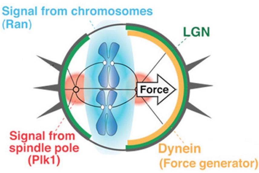
Diagram of how chromosomes are aligned
Courtesy of Nature Cell Biology and T. Kiyomitsu and I. Cheesman.
CAMBRIDGE, Mass. – To solve a mystery, sometimes a great detective need only study the clues in front of him. Like Agatha Christie’s Hercule Poirot and Arthur Conan Doyle’s Sherlock Holmes, Tomomi Kiyomitsu used his keen powers of observation to solve a puzzle that had mystified researchers for years: in a cell undergoing mitotic cell division, what internal signals cause its chromosomes to align on a center axis?
“People have been looking at these proteins and players in mitosis for decades, and no one ever saw what Tomomi observed,” says Whitehead Institute Member Iain Cheeseman. “And it’s very clear that these things are happening. These are very strong regulatory paradigms that are setting down these cell division axes. And careful cell biology allowed him to see that this was occurring. People have been looking at this for a long time, but never with the careful eyes he brought to it.”
Kiyomitsu, a postdoctoral researcher in Cheeseman’s lab, published his work in this week’s issue of the journal Nature Cell Biology.
The process of mitotic cell division has been studied intensely for more than 50 years. Using fluorescence microscopy, today’s scientists can see the tug-of-war cells undergo as they move through mitosis. Thread-like proteins, called microtubules, extend from one of two spindle poles on either side of the cell and attempt to latch onto the duplicated chromosomes. This entire “spindle” structure acts to physically distribute the chromosomes, but it is not free floating in the cell. In addition to microtubules from both spindle poles that attach to all of the chromosomes, astral microtubules that are connected to the cell cortex—a protein layer lining the cell membrane—act to pull the spindle poles back and forth within the cell until the spindle and chromosomes align down the center axis of the cell. Then the microtubules tear the duplicated chromosomes in half, so that ultimately one copy of each chromosome ends up in each of the new daughter cells.
The process of mitosis is extremely precise; when it comes to manipulating DNA, cells verge on being obsessive and with good reason. Gaining or losing a chromosome during cell division can lead to cell death, developmental disorders, or cancer.
As Kiyomitsu watched mitosis unfold in symmetrically dividing human cells, he noticed that when the spindle oscillates toward the cell’s center, a partial halo of the protein dynein lines the cell cortex on the side farther away from the spindle. As the spindle swings to the left, dynein appears on the right, but when the spindle swing to the right, dynein vanishes and reappears on the left side.
For Kiyomitsu, the key to the alignment mystery was dynein, which is known as a motor protein that “walks” molecular cargoes along microtubules. Kiyomitsu determined that in this case, dynein is anchored to the cell cortex by a complex that includes the protein LGN, short for leucine-glycine-asparagine-enriched protein. Instead of moving along an astral microtubule, the stationary dynein acts as a winch to pull on the spindle pole, and the microtubules and chromosomes attached to it, toward the cell cortex.
Kiyomitsu found that when a spindle pole comes within close proximity to the cell cortex, a signal from a protein called Polo-like kinase 1 (Plk1) emanates from the spindle pole, knocking dynein off of LGN and the cell cortex, stopping the spindle pole’s forward motion, and freeing dynein to move to the opposite side of the cell. These oscillations continue with decreasing amplitude until the spindle settles along the cell’s center axis.
As he was deciphering dynein’s role in spindle alignment, Kiyomitsu noticed that a layer of LGN extends all around the cell cortex, except in the areas that are closest to the chromosomes. As the chromosomes swing back and forth, the area cleared of LGN changes in response. Because dynein needs to anchor to LGN, this cleared area ensures that dynein can only attach and pull to the right and left of the aligning chromosomes, rather than from above and below.
After testing a couple of signaling molecules associated with chromosomes, Kiyomitsu determined that a signal from the chromosomes, involving the ras-related nuclear protein (Ran), blocks LGN, and therefore dynein, from attaching to the cell cortex closest to the chromosomes. Ran bound to guanosine-5'-triphosphate (Ran-GTP), which controls nuclear import in the interphase stage of mitosis, had previously been suggested to control spindle assembly during mitosis in germ cells, but roles for the Ran gradient in mitotic non-germ cells were unclear. Kiyomitsu’s work suggests a key role for Ran in directing spindle orientation.
Kiyomitsu says the axis that the spindle poles travel along is crucial to cells.
“The spindle orientation is critical for maintaining the balance between stem cells and mature cells during development,” he notes. “And if this orientation becomes dysregulated or misregulated, it is reported that this may contribute to causing cancer even if chromosomes are properly segregated.”
This work was supported by the Massachusetts Life Sciences Center, the Searle Scholars Program, and the Human Frontiers Science Foundation, the National Institutes of Health (NIH)/National Institute of General Medical Sciences, and the American Cancer Society.
* * *
Iain Cheeseman’s primary affiliation is with Whitehead Institute for Biomedical Research, where his laboratory is located and all his research is conducted. He is also an assistant professor of biology at Massachusetts Institute of Technology.
* * *
Full Citation:
Kiyomitsu, T., & Cheeseman, I. M. (2012). Chromosome- and spindle-pole-derived signals generate an intrinsic code for spindle position and orientation. Nature Cell Biology, 14(3), 311-317. doi:10.1038/ncb2440
Contact
Communications and Public Affairs
Phone: 617-452-4630
Email: newsroom@wi.mit.edu


