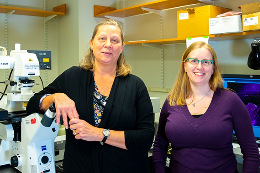
Lab Manager and Electron Microscopy Specialist
Nicki Watson (left) and Light Microscopy Specialist
Wendy Salmon (right)
Conor Gearin/Whitehead Institute
Nicki Watson and Wendy Salmon: W. M. Keck Microscopy Facility
Light Microscopy Specialist Wendy Salmon sometimes finds herself looking into microscopy images like shapes in the clouds. “I’ll say, ‘Look, there’s a snowman!’” she says. The habit comes a deep familiarity with the odd shapes found in microscopy, she says.
Lab Manager and Electron Microscopy Specialist Nicki Watson explains that these imaginative descriptions help communicate with researchers while exploring their images in the W. M. Keck Microscopy Facility. “If you looking through the oculars of the microscope, and you’re trying to describe what you’re seeing to someone who’s not looking, you don’t know if you’re looking at the same thing,” Watson says. “You get creative in trying to describe what you’re seeing.”
Since it takes a long time to learn electron microscopy techniques, Watson is able to save researchers time. “If they only need 3 or 4 pictures for one paper, it’s not worth their six months to train to get competent enough to produce publication-quality work,” she says. However, if someone’s paper or thesis focuses on electron microscopy, she helps train them on the equipment.
Similarly, Salmon tries to get researchers as independent as they can be if their light microscopy protocols are relatively straightforward. “It also makes them much more adept at navigating the results of their experiments,” Salmon says. “When they understand the instrumentation better, they can interpret what they’re seeing and make decisions on their own.”
Watson recalls a particularly satisfying project in which she achieved something she did not think was possible. A researcher was looking for a tiny developing kidney in an embryonic zebrafish, but was unsure what it would look like — making it difficult to locate. Doubtful at first, Watson searched through section after section. “I kept at it, looking and looking, and finally saw something,” she says. “Not surprisingly, it looks like a kidney, with glomeruli and tubules.”
Sometimes, microscopy can reveal surprises. Salmon once assisted researchers trying to make time-lapse images of mammary organoids to see if the organoid’s structure would change over time. When the team played back the overnight time-lapse, they saw something unexpected — cells were roaming around within the larger structure, and the video clearly captured their journeys.
Microscopy brings a unique perspective to research projects, Salmon says. “Almost everything else is a bulk measurement of some sort,” she says. “With microscopy you know where something is happening. You get spatial context.”
“That’s true right down to the ultrastructural level on the electron microscope, too,” Watson agrees. “In electron microscopy, you can look at how these perturbations are affecting the ultrastructure of the cell, at a high-resolution level.”
Topics
Contact
Communications and Public Affairs
Phone: 617-452-4630
Email: newsroom@wi.mit.edu


