Protein-printing technique gives snapshots of immune system defense

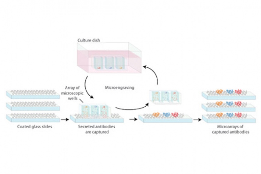
Researchers sorted individual immune cells into tiny wells in a microengraved rubber device. The cells then “printed” their antibodies onto glass slides. Multiple protein microarrays were created this way from the same set of B cells, allowing scientists to gather a much more complete picture of how immune cells react under attack.
Illustration: Tom DiCesare
CAMBRIDGE, Mass. — When Albrecht Durer and other Renaissance artists painstakingly etched images onto plates, swabbed ink into the fine grooves and transferred the images to paper with a press, they never could have guessed that centuries later the same technique would uncover the secrets of human cells.
Whitehead Institute and Massachusetts Institute of Technology researchers have borrowed a technique from such “intaglio” printing to create snapshots describing the behavior of immune cell populations at a moment in time.
The work may aid vaccine research and eventually lead to clinical devices that change the way physicians diagnose and treat infection and disease, suggests J. Christopher Love, a former postdoctoral associate in the lab of Whitehead Member Hidde Ploegh and now an MIT assistant professor of chemical engineering.
|
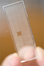
J. Christopher Love, Craig Story, Chih-Chi Andrew Hu and Eliseo Papa designed and tested a system that “prints” the antibodies created by single immune cells onto a glass slide (below) coated with capture antibodies, producing protein microarrays. Photos: Whitehead
|
For the first time, the method developed by Love and his colleagues lets researchers look at single white blood cells and measure specific characteristics of the antibodies they produce when the body is under attack.
The white blood cells called B cells can offer billions of combinations of antibodies that bind to bacteria, viruses or toxins, flagging the invaders for destruction. “There are no two cells with the exact same signature,” notes Hidde Ploegh.
And neither is there much information about what combination of antibodies goes with what target, Ploegh adds. But isolating and keeping B cells alive long enough to investigate them is inefficient and time-consuming, and it has been impossible to fully correlate analyses of all the antibodies.
In recent years, biologists have begun to exploit a set of techniques, collectively called soft lithography, to transfer patterns of biological materials to microarrays, much as Renaissance artists transferred images to paper.
Love came up with a new twist on this idea: individual cells could make the ink.
To test the idea, Love, visiting scientist Craig Story and their colleagues put living mouse B cells into a microengraved rubber device like the tiniest and densest of ice cube trays. There, 20,000 cells, separated into individual compartments, churned out their unique brands of antibodies produced after a series of immunizations.
The researchers sealed the tiny chambers with microarrays—glass microscope slides coated with capture antibodies. The secreted proteins stuck to the glass, seeding it like a printer’s plate being imbued with ink.
Next, the scientists stamped out multiple copies of these microarrays. Studying these, the scientists can examine the antibodies expressed by a single cell, in the context of a whole population of cells. The information is extracted and integrated by customized software and the large sets of data are examined much as they are with DNA microarrays.
The first results, imaged and analyzed by graduate student Eliseo Papa of the Harvard/MIT Health Science and Technology Institute, appeared in PNAS online the week of November 3. They gave a snapshot of the diversity of B cells that were generated by a series of immunizations designed to mimic a multi-part vaccination.
This technique could prove a powerful tool for research and medical testing. Currently, “the only accepted measure of immunological protection is a relatively crude test for whether antibodies are present in the blood,” Ploegh says. “Knowing not only the level of antibody in a patient’s blood but also how effective the antibodies would be in fighting a specific disease would be a big step.”
Knowledge gained this way would be vastly helpful for vaccine development. “It’s extremely important to have a rapid test to interrogate a vaccine recipient’s immune system,” allowing widespread testing among a given human population, says Ploegh.
“Nobody really knows in detail what happens between vaccine boosters,” adds Papa. “Multiple booster injections are given irrespective of the actual response.” If a cost-effective, portable device could work on this method, it could help gauge the progression of vaccine boosters or infectious illnesses on the basis of patients’ real-time immune responses, and help doctors weigh treatment options based on those responses.
Moreover, Love says, further development of the technique could make it possible to quickly determine whether an infection is bacterial or viral, figure whether a cancer patient has built up enough “immunity” to his or her cancer to forgo another round of immunotherapy, and obtain complete profiles of an immune response with as little as a drop of blood—a crucial ability when testing infants and children.
* * *
Hidde Ploegh’s primary affiliation is with Whitehead Institute for Biomedical Research, where his laboratory is located and all his research is conducted. Ploegh is also a professor of biology at Massachusetts Institute of Technology.
* * *
Story, C. M., Papa, E., Hu, C. A., Ronan, J. L., Herlihy, K., Ploegh, H. L., & Love, J. C. (2008). Profiling antibody responses by multiparametric analysis of primary B cells. Proceedings of the National Academy of Sciences, 105(46), 17902-17907. doi:10.1073/pnas.0805470105
Topics
Contact
Communications and Public Affairs
Phone: 617-452-4630
Email: newsroom@wi.mit.edu

