
Whitehead Institute postdoctoral researcher Ashley Bonneau
Lucila Scimone/Whitehead Institute
Meet a Whitehead Postdoc: Ashley Bonneau
Ashley Bonneau is a postdoc in Whitehead Institute Member Peter Reddien’s lab investigating signaling pathways involved in embryonic development and tissue regeneration. We sat down with Bonneau to learn more about her and her experiences in and out of the lab.
What species do you work with in your research?
The main species that we work with in the Reddien lab is the planarian Schmidtea mediterranea, a small flatworm with a remarkable and robust ability to undergo whole body regeneration. If you cut one into ten pieces, you get ten worms. They're a wonderful model to understand the process of how new cells are established and how they're integrated across a multicellular animal. They have many complex tissue types, including a nervous system, muscle, and a digestive and excretory system. If you cut off or remove any of those parts, they're able to fully replace them.
What do you investigate?
The lab’s main focus is understanding how different cell types, and therefore different tissues, are established, integrated, and maintained during regeneration and normal tissue turnover. During embryonic development, cells and tissues develop in a linear progression from a set starting point. Whereas during regeneration your starting point is always going to be different because each injury, and therefore tissue missing, is going to be unique. The question then is how does the animal know what’s missing, what tissue to make, and how to integrate it into the existing tissues.
One of the key findings has been that there are highly conserved signaling pathways that predominantly regulate this process, providing instructions to guide regeneration. Many of the genes involved are the same ones that are required to specify different cell fates—the final identity or type of each cell—during development. Remarkably in the adult, this patterning information is harbored in mainly one cell type: muscle. Coming from a developmental biology background, I am interested in understanding when and how this patterning information is refined and restricted.
When you were a kid, what did you want to be when you grew up?
In elementary school I wanted to be a lawyer. I really enjoyed arguing with my sister and I thought that it would be fantastic as a career. Then in high school I became fascinated with science. Initially I was interested in becoming an archaeologist but eventually I transitioned to molecular and cellular biology.
What led you to your current area of research?
I did my undergraduate work at Cal State University Channel Islands. It's a very small school but it had a really great focus on student mentoring and undergraduate research. From there I went to Yale and completed my PhD under the direction of Antonio Giraldez. The majority of my work as a PhD student was aimed at identifying how the genome is activated during development. It was during my graduate work that I become interested in patterning and how cell fate decisions are made. I was fortunate to attend a talk given by Peter during my first year in the program. I became interested in both the model and the types of questions the Reddien lab was focused on investigating.
What are the most and least fun parts of doing research?
The most fun part is you get to continuously ask questions that no one else in the world knows the answer to, and you actually (hopefully) get to provide that answer. A fantastic aspect of being a scientist is getting to discover the unknown and sharing that information with the community. On the flipside, because you’re working with the unknown, if you're having issues or things aren't working, it's really hard to troubleshoot. For every successful experiment you might have had ten failures. As a scientist you really need to develop the skill of moving past failures so that you can get to the success stories. It's a big challenge but also part of the fun.
What’s the biggest disaster you’ve ever had in the lab?
During my PhD, I worked with a lot of radioactivity. I was teaching a new post-bac student how to work with it safely and I set down the glass cylinder that holds the radioactive material to explain something to her. I didn't realize the cylinder had tipped and started to roll, and it rolled and shattered on the floor. Obviously, we had procedures in place, we were prepared, but I mean I was training someone on how to be safe with radioactivity and then I had this spill. It was not an ideal situation, but I was able to turn it around and make it a teaching moment about what to do in that situation.
What are your hobbies or passions outside of work?
I'm a mom so I'm very passionate about raising my daughter and making her excited about the world. I'm also a newborn and family photographer. I took photography classes in high school and I found the creative process really satisfying. Then when my daughter was born, I purchased a hobbyist digital camera to record special memories. I found myself taking over a hundred pictures a day and fell in love with capturing different moments of her childhood. I have only recently branched out into taking photos for other families. I like to focus on the candid aspects of a session, capturing true memories for people.
I also own a small children's online boutique. I make stuffed animals, bonnets and hats for children, and lovies for newborn babies. Again, I was inspired by my daughter. For her first Halloween I delayed ordering her a Cabbage Patch wig and could not purchase one in time. After many YouTube tutorials, I taught myself to crochet in order to make her one. After that, I wanted her to have handmade, keepsake toys and picked up sewing. That snowballed into a children's store.
How much of what you make do you keep?
All of my new prototype items go to my daughter. She gets all the misfits and the maybe not perfectly sewn things. She has more stuffed animals than any other four-year-old that I know.
What’s the most visually striking thing you’ve seen in the lab?
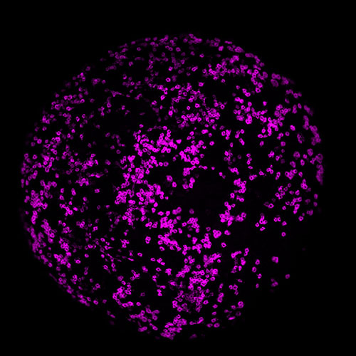
Planarian embryos have a dividing population of cells called neoblasts (stem cells) that will eventually give rise to all the cell types in the animal. When I first got my florescent probes to work, I could see the neoblast distribution and pattern as the embryo develops. They look like beautiful little planets and it’s quite striking.
Where do you see yourself in ten years?
I hope to be teaching at a smaller liberal arts college and leading my own research team. Coming from an undergraduate institute that was focused on mentoring undergraduate research, I hope to integrate that into my future lab as well.
Contact
Communications and Public Affairs
Phone: 617-452-4630
Email: newsroom@wi.mit.edu


