In regenerating planarians, muscle cells provide more than heavy lifting

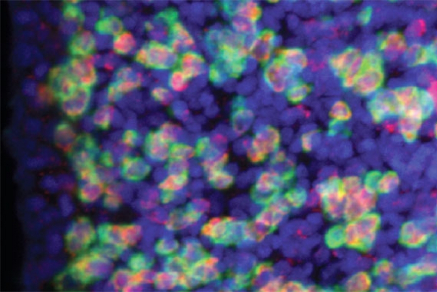
Planarian muscle cells expressing 19 position control genes
Courtesy of Cell Reports
CAMBRIDGE, Mass.– By studying the planarian flatworm, a master of regenerating missing tissue and repairing wounds, Whitehead Institute Member Peter Reddien and his lab have identified an unexpected source of position instruction: the muscle cells in the planarian body wall.
“I was completely surprised. We had no idea it would be muscle,” says Reddien, who is also an associate professor of biology at MIT. “Finding such a cellular system for positional control in an adult regenerative animal was unanticipated and is very informative for understanding regeneration.”
For decades, researchers studying regeneration have focused on stem cells, largely because of their ability to spawn almost any type of replacement cell. In fact, the Reddien lab recently determined that cells known as cNeoblasts are the planarian stem cells that are able to regenerate all tissues in these animals.
Long neglected, however, has been the question of cell identity: how do cNeoblasts know where they are and which cells are needed to recreate missing tissues?
The Reddien lab and other labs had already identified various planarian genes involved in imparting positional information during regeneration and adult tissue maintenance, including regulators of Wnt, Bmp, and Fgf signaling pathways. These position control genes (PCGs) are highly conserved and many are found in other animals, including humans. But the cellular location of where those signals originated was unknown.
Reddien hypothesized that a certain type of cell expresses PCGs, possibly a unique type of cell that had not yet been described. To identify these cells, Jessica Witchley and Mirjam Mayer studied PCG expression in planarians and determined that all tested PCGs are indeed expressed in the same cells. They found that collagen, a telltale marker of planarian muscle cells, is also expressed in the same cell population. When part of a planarian was cut off, the muscle cells in the body wall altered which PCGs they expressed in response to the wounding. The lab’s results are described in the August 29 issue of Cell Reports.
Until now, the planarian’s muscles in the body wall were thought to primarily allow the worms to twist, turn, and respond to touch, in addition to providing some structure to their boneless bodies. Yet, these functions may also explain why muscle cells are so well suited to secrete the proteins coded for by the PCGs.
“Because the muscle cells extend throughout the organism, from head to tail, they are a great way to influence delivery of the PCGs’ proteins and get signaling throughout the animal,” says Witchley, a co-first author of the Cell Reports paper and a former technician in the Reddien lab. “Because the muscle fibers are long, when you cut the animal, it can immediately respond by contracting to close the wound, turning on wound-induced genes at that site, and eventually changing the pattern of expression to respond to the missing tissue.”
The discovery of muscle cells’ remarkable role in regeneration opens whole new areas of research.
“For one, we’d like to know how information from muscle cells at the wound site is actually conveyed to the stem cells and how the stem cells receive that information and respond,” says Mayer, co-first author and former postdoctoral researcher in the Reddien lab. “And it would be very interesting to see if muscle cells have a position control role in other animals that is similar to what we’ve found in planarians.”
This work is supported by the National Institutes of Health (NIH grant R01GM080639), the Keck Foundation, and the Helen Hay Whitney Foundation.
* * *
Peter Reddien’s primary affiliation is with Whitehead Institute for Biomedical Research, where his laboratory is located and all his research is conducted. He is also a Howard Hughes Medical Institute Investigator and an associate professor of biology at Massachusetts Institute of Technology.
* * *
Citation:
Witchley, J. N., Mayer, M., Wagner, D. E., Owen, J. H., & Reddien, P. W. (2013). Muscle cells provide instructions for planarian regeneration. Cell Reports, 4(4), 633-641.
Contact
Communications and Public Affairs
Phone: 617-452-4630
Email: newsroom@wi.mit.edu


