Emerald City: How a jellyfish helped advance science
CAMBRIDGE, Mass. — Salmon fishermen trolling along the waters off Puget Sound in Washington are often witness to an awesome sight when they haul in their catch: salmon captured in nets that glow brilliantly against the nighttime sky. This unnatural sheen is caused by the Northwest Pacific jellyfish Aequorea victoria, whose cellular makeup includes a bioluminescent protein called aequorin that a emits a deep blue light. Another protein called green fluorescent protein, or GFP, absorbs this light, and through a biophysical process, turns it into a glowing emerald green.
The jellyfish has given off its evening shine for centuries, but in the early 1990s, scientists developed a way to use GFP in biological studies. Researchers removed the gene for GFP from jellyfish, cloned it, and introduced it into the cells of the bacterium E. coli and in C. elegans, a soil nematode widely used as a biological model. In the experiments, GFP clones were fused to specific proteins in the study models, causing those proteins to glow when illuminated with blue light. Under a microscope, these GFP-labeled proteins shine in a steady glow, allowing scientists to easily track and observe the proteins’ movements and behaviors. Some 10,000 studies a year now report findings related to GFP’s use in the laboratory. GFP has even been used to make “transgenic art,” notably a fluorescent rabbit named Alba, who was bred in 2001 with this jellyfish protein imbedded into her genetic code.
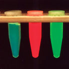 Glowing samples of GFPs Image: William Ward
Structurally, GFP looks like a barrel that surrounds a light bulb in its interior. The interior bulb, which is known technically as a chromophore, doesn’t produce its own light, but rather absorbs and alters light from another source. In a jellyfish, that source is the aequorin protein, which transfers its blue light to GFP by a quantum process that some scientists compare to mind-reading—unlike sound waves, energy moves from one molecule to the other in the absence of any medium. In the laboratory, artificial light substitutes for aequorin’s natural role.
Today, scientists use GFP to determine where proteins are located during different stages of a cell’s life, or to watch how proteins interact to produce disease. GFP also helps scientists track the introduction of foreign genes into DNA, a strategy used to create transgenic models to study cancer, diabetes, and other diseases.
GFP one day could play a role in treating illnesses like cancer. Scientists hope to be able to incorporate GFP directly into tumor cells, causing them to glow as a distinct population, easily separated from healthy cells and tissues. Ultimately, GFP use in research will only grow. The little green protein looks to have a bright—indeed a glowing—future in biomedical research.
Topics
Contact
Communications and Public Affairs
Phone: 617-452-4630
Email: newsroom@wi.mit.edu


